


Hi! - ENG


The use of healing abutments during the treatment enables us to preserve the tissue architecture, reduce the treatment time and provide a good esthetic result.
INTRODUCTION
The placement of implants in post-extraction regenerated alveoli is a treatment with good results in terms of tissue healing and survival, which the literature associates with few complications.
The key factors for success in this type of treatment are based on a series of clinical principles such as atraumatic tooth extraction to preserve the alveolar bone, the absence of an active infection and primary stability of the implant.
The use of healing abutments during the treatment enables us to preserve the tissue architecture, reduce the treatment time and provide a good esthetic result.
KEYWORDS: alveolar ridge maintenance, emergency profile, soft tissue, PEEK abutment
DESCRIPTION OF THE CLINICAL CASE
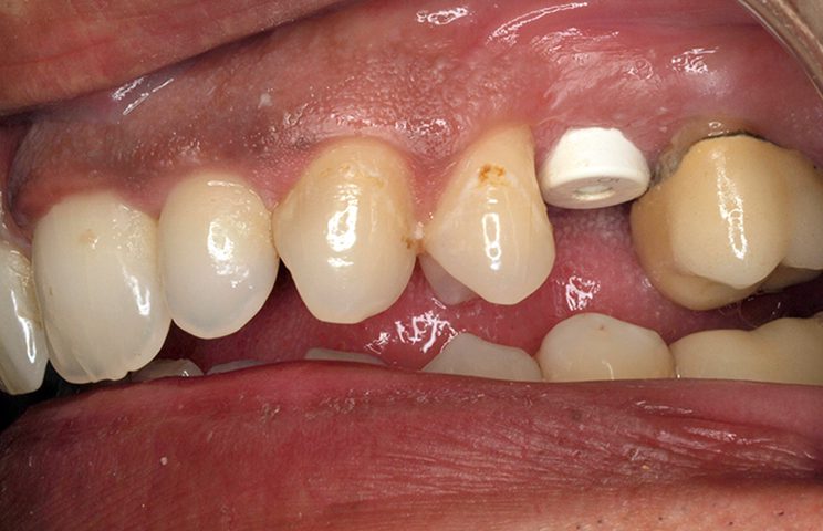
A 40-year-old male patient who smokes 15 cigarettes per day with no medical pathology or history of interest presents at the office due to a loss of a pin-retained crown on position 25 with no possibility of restoration. Anamnesis was conducted and clinical and radiological examinations were performed on the remaining root.
![]()
With the patient’s consent, we decided to extract the remaining root to place an implant there and to restore the tooth with a screwed metal-ceramic crown in a second phase. On the same day of surgery, we decided to work the soft tissue profile with a PEEK healing abutment.
Surgical technique
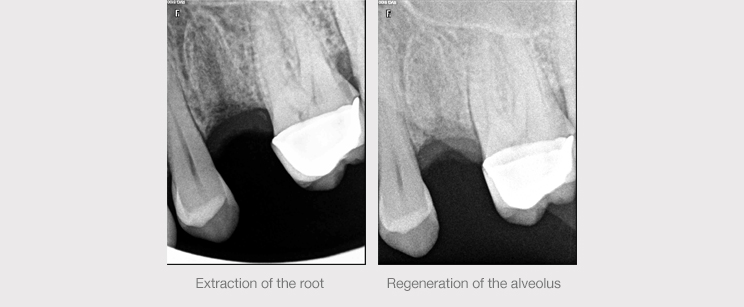
Examination of the bone architecture via CAT scan revealed a fine vestibular table, a very wide alveolus in the vestibular-palatine sense and very little apical bone height towards the maxillary sinus, which is a very important area for achieving primary stability. This is why we decided to conduct the treatment in two phases, with an extraction of the remaining root and an alveolar ridge maintenance technique followed by surgery to place the implant in the regenerated alveolus.
Extraction of the root and careful curettage of the area were performed. This was followed by guided alveolar regeneration with collagenated heterologous bone and pericardium membrane. Maintenance of the alveolar ridge via regeneration techniques helped to minimize residual resorption and consequent soft tissue collapse, which would have produced unacceptable esthetic results in prosthesis on implant.
After a three-month healing period, we proceeded to place a Biomimetic Ocean CC implant measuring 3.5 x 11.5 mm.
Choosing a conical connection provides many advantages. The design of the connection makes it possible to evenly distribute stresses and reduce stress peaks away from the ridge bone area. The narrowing of the micro-gap, together with a larger support surface at the abutment-implant interface, provides mechanical advantages such as a reduction in micro-movement and biological benefits like the minimization of bacterial leaks. For all these reasons, we considered it best to use this type of connection to enable a tight soft tissue seal and guarantee optimum esthetic results in the long term.
![]()
Treatment of the tissues
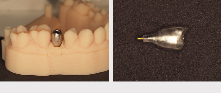
We decided to wait three months to achieve a good osseointegration of the implant. In the second surgical operation, we placed a PEEK healing abutment (Avinent Implant System) with an anatomical shape adapted for molars that was easy to adjust in the mouth for purposes of customization and that could optimize the emergency profile of the tissues.
This innovative material is a good alternative to metal alloys that expands the possibilities of treatment and obtains very good results. It stands out for being inert and not cytotoxic, presenting low thermal and electrical conduction and lowering the adhesion of bacterial plaque.
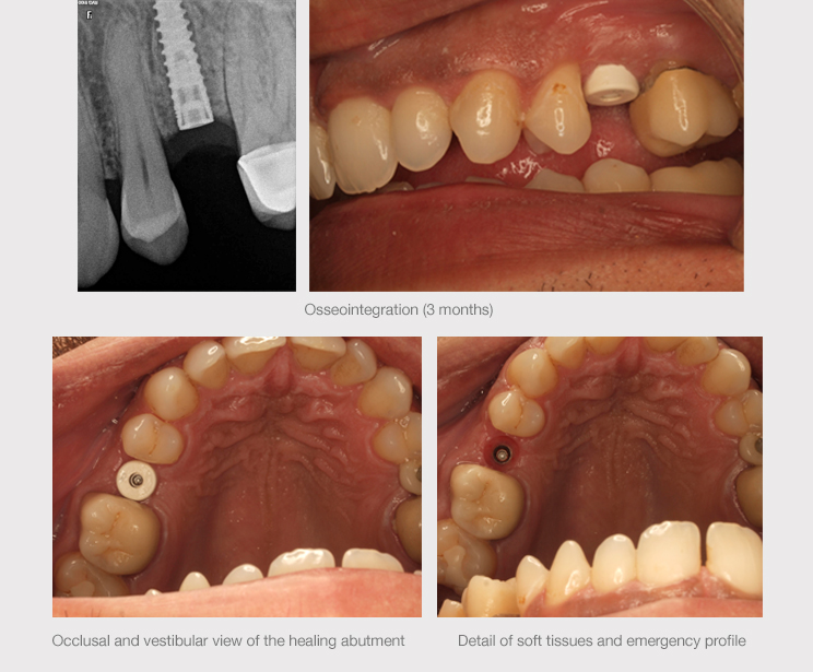
During this second operation, we also increased the volume of the vestibular peri-implant mucosa using the roll technique. The images below show the fully healthy state of the tissues and the volume increase achieved with this technique, together with the placement of the shaper in the second operation.
Definitive restoration
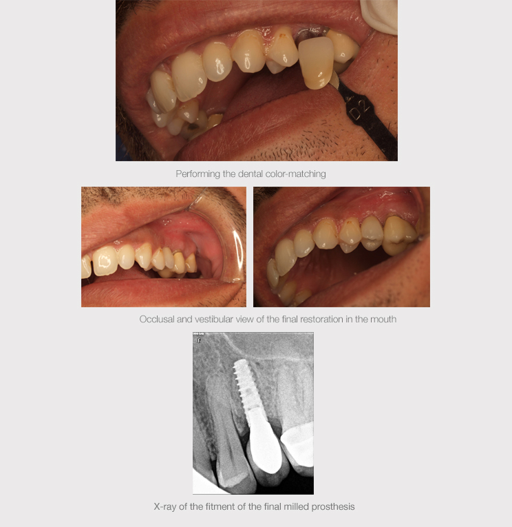
Three weeks later, once the desired emergency profile was achieved, the scanbody was screwed to the implant to take a digital impression with the 3Shape TRIOS intraoral scanner and to be able to design the final restoration.
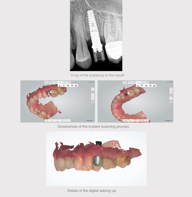
The scanned intraoral file was sent to the prosthetic laboratory to design the final crown. This design was then sent to the AVINENT milling center where it was milled in CoCr. A 3D working model of the patient’s mouth was also created with additive manufacturing technology.
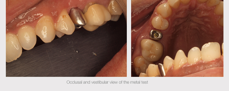
During the metal test, the prosthesis was verified to fit perfectly in the mouth and dental color-matching was performed so that the prosthetic laboratory could finish the restoration by loading the ceramic.
CONCLUSION
Implant placement in post-extraction regenerated alveoli is currently a widely described treatment with a high success rate in terms of both tissue healing and survival.
The long-term success of any implant treatment will depend on biological, mechanical, functional and esthetic criteria and conical connections may be a good choice to meet these criteria.
In addition, the development of new materials with biocompatible and biomechanical properties like PEEK allows us to expand the possibilities of treatment, attaining optimal results.